A human clinical trial using ultrasound and microbubbles to enhance gemcitabine treatment of inoperable pancreatic cancer
Georg Dimcevskia, Spiros Kotopoulisa, Tormod Bjånesc, Dag Hoemd, Jan Schjøttc, Bjørn Tore Gjertsenf, Martin Biermannh, Anders Molveni, Halfdan Sorbyek, Emmet McCormacke, Michiel Postemal, Odd Helge Giljaa, Journal of controlled release : official journal of the Controlled Release Society. okt. 2016.
Diagnostic ultrasound imaging is commonly used throughout the clinic in a multitude of fields such as cardiology, gastroenterology, gynaecology, etc. To improve the signal to noise ratio and help visualise tissue perfusion stabilised gas microbubbles (2–10µm in diameter) are injected into the blood stream. These are considered biologically inert, and degrade after several minutes in the body.
In vitro and pre-clinical work has shown that by modifying the ultrasound emission conditions and co-injecting a chemotherapeutic agent with the microbubbles can significantly improve the efficacy of the therapeutic agent. This technique is commonly known as sonoporation. This increased efficacy is due to the local interaction between the microbubbles and cells, increasing the cellular porosity and potentially the vascular permeability. A benefit of this technique is that this effect only occurs where there is ultrasound and microbubbles present resulting in a non-invasive, image-guided drug delivery technique.
Researchers from the Department of Clinical Medicine (K1), along with collaborators, have just published the completed results of the world first clinical trial evaluating the toxicity and efficacy of sonoporation on patients with pancreatic ductal adenocarcinoma. Patients were treated using the standard chemotherapeutic regimen (Gemcitabine infused over 30 min). Immediately following completion of the infusion, microbubbles were injected every 3.5 minutes and ultrasound was applied to the tumour.
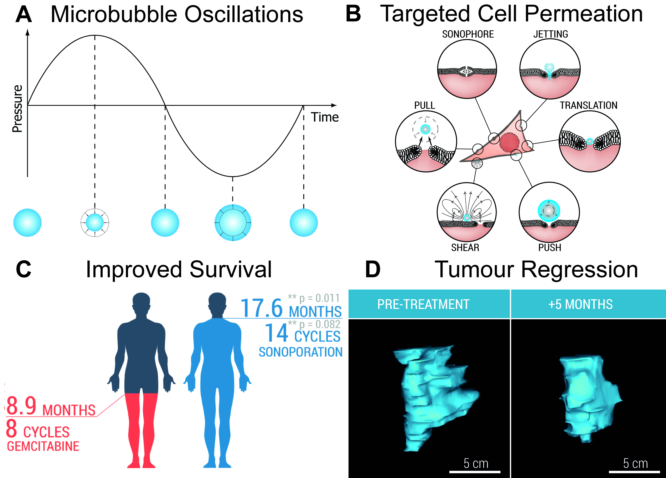
After 138 treatment cycles, over 10 patients, no increase in overall toxicity was observed. Patients were able to undergo more than double the number of treatment cycles when comparing to the chemotherapy treatment alone (8.3 ± 6.0 cycles, versus 13.8 ± 5.6 cycles) indicating an extended period of high-quality of life, whilst in 50% of the patients’ tumour size reduction was observed. In addition, median survival was almost doubled, from 8.9 to 17.6 months.
 Caption: Ultrasound waves makes microbubbles resonate (Panel A), which induces numerous interactions between the microbubbles and cells (Panel B). When performed in conjunction with chemotherapy, survival and number of treatment cycles increase (Panel C). Tumour volume regression is also observed in several patients (Panel D).
